
The ability to turn genes on or off is central to the diversity we see in cells, individuals, and even in terms of health and disease. This process, known as gene transcription, involves converting the information stored in our DNA into a carbon copy called RNA. Until recently, scientists have relied on inaccurate illustrations and indirect experiments to understand this process, as it occurs at the molecular level and is not directly visible. However, a revolutionary microscopy technique now allows researchers to observe never-before-seen molecular processes within genetic material, providing valuable insights into how genes are switched on and regulated.
Antonio Giraldez, PhD, Fergus F. Wallace, professor of genetics at Yale School of Medicine, studies the DNA codes in the genome and how cells interpret these codes to create an embryo. A crucial aspect of understanding these processes involves our ability to visualize the genome. Unfortunately, traditional microscopy methods have limitations. To overcome these limitations, Giraldez and his colleagues, including first author of the studies, doctoral student Mark Pownall, collaborated with Joerg Bewersdorf, PhD, Harvey and Kate Cushing professor of cell biology and professor of biomedical engineering and physics, as well as a renowned microscopy expert, to develop a new technique called chromatin expansion microscopy (ChromExM).
In an article published online in Science on July 6 demonstrate its success in increasing the physical volume of zebrafish embryonic cell nuclei 4,000-fold to dramatically improve image resolution. The technique has allowed researchers to see for the first time how single molecules shape gene expression in cells during embryonic development and to come up with a new model of how genes are regulated.
Our research allows us to see the fundamental processes in the nucleus that underlie everything in life, from the formation of an embryo to cancer, Giraldez says. We can see processes that we could only imagine before.
Our research allows us to see fundamental processes in the nucleus that underlie everything in life, from the formation of an embryo to cancer. We can see processes that we could only imagine before.
Antonio Giraldez, Ph.D
After sperm fertilizes an egg, the genome is initially silent, Giraldez says. The fertilized egg must develop into a transient pluripotent stem cell, or a cell that can give rise to many different cell types, to develop into a healthy embryo. Programming this cell’s ability to make other cells requires jumpstarting the genome.
For years, Giraldez and his team have studied how the genome is activated. They’ve come a long way, from identifying key players to learning which genes are turned on. But we’d never seen the genome activate for ourselves, Giraldez says. There is a difference between describing how things might happen and actually witnessing how things are happening.
ChromExM microscopy technique helps researchers visualize the genome
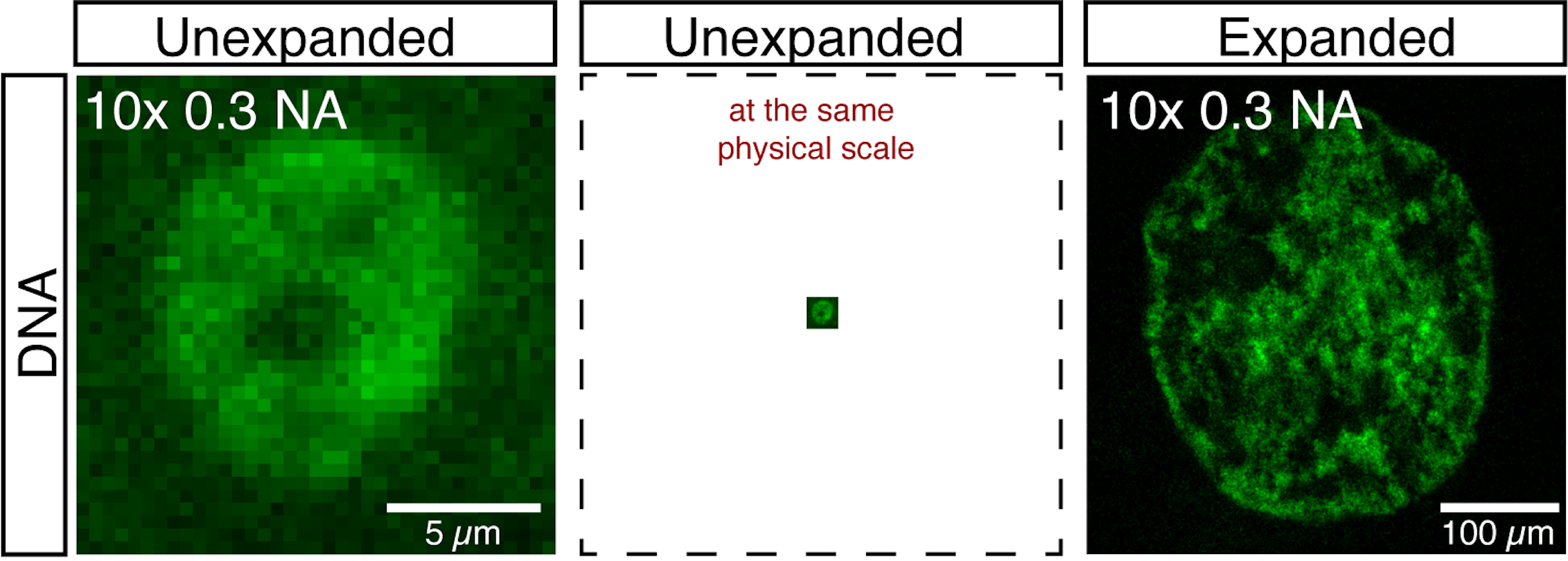
In his earlier work, study co-senior author Bewersdorf developed a technique called pan-ExM that involved anchoring cells to an expandable gel to allow visualization of cellular features with unprecedented resolution. As the gel expanded, it separated the cell and the proteins within them while maintaining their spatial organization, until the cell was 64 times larger in volume. Then, with a second gel, the team repeated the process so that the cell volume grew 4,000-fold. For this new study, the Giraldez and Bewersdorf labs teamed up to create ChromExM and applied it to embryos to visualize how genes are regulated. Now, each individual cell was roughly the size of an embryo. We used a very conventional instrument, a confocal microscope, which allowed us to get this incredible resolution of the molecular machinery of the cell when combined with ChromExM, Giraldez says. Even the most powerful microscopes could not view it.
The process, she explains, is like toy eggs expanding into dinosaurs when placed in water. When the egg is first dropped into the glass, the dinosaur’s features are not yet visible. But as the toy grows, it transforms from something amorphous into a creature with detailed features. That dinosaur probably grew two or three times larger, Giraldez says. Now he imagines that growth on a 4,000-fold scale.
Through ChromExM, the team was able to see fundamental genome processes in action for the first time. This allowed them to develop a new model of how genes are regulated, which they called kiss-and-kick to describe the transience of how regulatory regions in DNA called enhancers interact with gene initiators (promoters) to trigger the expression of the gene and how the transcript burst separates the regulatory regions of the gene (or kicks out the enhancer) to pause expression. It’s like going from the pixelated black-and-white cell phone screens of the 1980s to today’s big, colorful, ultra-high-definition screens, says Giraldez. Our technique allowed us to see details that weren’t possible before.
With this new method, the team is eager to test hypotheses that were untestable until recently. For example, as well as seeing fundamental molecular processes, they also hope to explore how different genes are turned on or off, how they are positioned relative to other genes in the nucleus, and how mutations affect the positions of genes. Also, while other microscopy techniques may be prohibitively expensive, ChromExM is accessible to most laboratories. Our work will democratize a method for seeing how molecular processes occur inside the nucleus, which will open up new areas of research, Giraldez says.
The team now hopes to further improve the resolution of its technique. Although researchers are now able to visualize molecules interacting with the genome, they are not yet able to identify individual genes. Imagine you are in space and take a picture of New York City. Before, you could only see the island, but now you can see the people in the city, Giraldez explains. But we still don’t know who these people are. If you think of those people as the geniuses we want to see, then we want a camera that allows us to focus on individual people. This detail will allow scientists to understand the fundamental principles of how genes are turned on and off, broken or repaired, and how mutations affect their functional steps critical to understanding how our genes function in health and disease.
#Supersize #Cell #unlocking #secrets #genome #expansion #microscopy
Image Source : medicine.yale.edu